Odovtos-International Journal of Dental Sciences (Odovtos-Int. J. Dent. Sc.), Online First, 2025. ISSN: 2215-3411
https://doi.org/10.15517/caks2s94

https://revistas.ucr.ac.cr/index.php/Odontos
BASIC RESEARCH:
Cleaning Protocols for Bioceramic & Epoxy Resin Endodontic Sealers:
Adhesion and Residue Removal
Protocolos de limpieza para cementos endodónticos biocerámico y a base de resina epóxica:
adhesión y eliminación de residuos
Jaime Barcena-Taco¹ https://orcid.org/0000-0003-3082-3369
Ivana D. Mamani-Colque¹ https://orcid.org/0000-0001-6991-2612
Eliam C. Zúñiga-Mayhua² https://orcid.org/0009-0004-7621-6227
Wilfredo G. Escalante-Otárola² https://orcid.org/0000-0003-4879-3938
Milton C. Kuga3 https://orcid.org/0000-0001-5728-8293
Gabriela M. Castro-Núñez² https://orcid.org/0000-0002-2586-541X
¹School of Dentistry, Faculty of Health Sciences, Jorge Basadre Grohmann National University - UNJBG, Tacna, Peru.
²School of Dentistry, Universidad Catolica de Santa Maria - UCSM, Arequipa, Peru.
3Department of Restorative Dentistry, School of Dentistry, Araraquara, São Paulo State University (UNESP), Araraquara, SP, Brazil.
Correspondence to: Wilfredo Gustavo Escalante Otárola - wilfredo.escalante@ucsm.edu.pe
Received: 15-V-2025 Accepted: 19-IX-2025
ABSTRACT: This study evaluated the effect of different chemo-mechanical cleaning protocols on the residual presence of epoxy resin-based and bioceramic endodontic sealers, as well as the bond strength of a universal adhesive system for cementing fiber posts to intraradicular dentin. A total of 160 bovine incisors were prepared using the ProTaper system and filled with either an epoxy resin-based sealer (Vioseal) or a bioceramic sealer (BioRoot RCS). Four cleaning protocols were tested: ethanol (ET), amyl acetate (AA), acetone (AC), and an experimental solution (EX) combining amyl acetate, ethanol, and acetone. Residual sealer was analyzed via scanning electron microscopy, and bond strength was assessed through push-out tests. Fracture patterns were examined under a stereomicroscope, and data were analyzed using ANOVA and Kruskal-Wallis tests (α=5%). For epoxy resin-based sealers, AA and AC resulted in greater dentinal tubule opening than ET and EX (p<0.05), with AC leaving the least residue. Conversely, ET and EX showed higher residual presence. For bioceramic sealers, ET and EX achieved the greatest tubule opening (p<0.05). Bond strength was highest with ET in the apical third (epoxy sealer) and in the cervical third (bioceramic sealer) (p<0.05). Ethanol and the experimental solution effectively removed bioceramic sealer residues while preserving bond strength. Although epoxy resin-based sealers left more residue, all tested protocols provided satisfactory adhesion.
KEYWORDS: Intraradicular dentin; Bioceramic cement; Epoxy resin cement; Bond strength; Cleaning protocols.
RESUMEN: Este estudio evaluó el efecto de diferentes protocolos de limpieza quimico-mecánica sobre la presencia residual de cementos endodónticos a base de resina epóxica y biocerámico, así como la resistencia de unión de un sistema adhesivo universal para cementar postes de fibra a la dentina intraradicular. Se utilizaron 160 incisivos bovinos que fueron preparados con el sistema ProTaper y obturados con un cemento a base de resina epóxica (Vioseal) o un cemento biocerámico (BioRoot RCS). Se probaron cuatro protocolos de limpieza: etanol (ET), acetato de amilo (AA), acetona (AC) y una solución experimental (EX) que combina acetato de amilo, etanol y acetona. El cemento residual fue analizado mediante microscopía electrónica de barrido, y la resistencia de unión se evaluó a través de pruebas de push-out. Los tipo de falla adhesiva se examinaron bajo un estereomicroscopio, y los datos se analizaron utilizando los tests ANOVA y Kruskal-Wallis (α=5%). Para los cementos a base de resina epóxica, AA y AC resultaron en una mayor apertura de los túbulos dentinarios que ET y EX (p<0.05), siendo AC el que dejó menos residuo. Por el contrario, ET y EX mostraron mayor presencia residual. Para los cementos biocerámicos, ET y EX lograron la mayor apertura de los túbulos (p<0.05). La resistencia de unión fue mayor con ET en el tercio apical (sellador epóxico) y en el tercio cervical (sellador biocerámico) (p<0.05). El etanol y la solución experimental eliminaron eficazmente los residuos del cemento biocerámico mientras preservaban la resistencia de unión. Aunque los selladores a base de resina epóxica dejaron más residuo, todos los protocolos probados proporcionaron una adhesión satisfactoria.
PALABRAS CLAVE: Dentina intraradicular; Cemento biocerámico; Cemento de resina epóxica; Resistencia de adhesión; Protocolos de limpieza.
INTRODUCCION
Epoxy resin-based endodontic sealers are well known for their excellent sealing properties within the root canal, which is crucial for the long-term success of endodontic treatment (1). Despite their moderate cytotoxicity, their clinical performance has proven to be biocompatible, reinforcing their popularity in therapeutic applications (2). This combination of effective sealing and biocompatibility has allowed epoxy resin-based sealers to remain a reliable choice in endodontics.
On the other hand, bioceramic endodontic sealers, primarily composed of calcium silicate (3), stand out not only for their sealing capabilities but also for their unique bioactivity (4,5), which promotes the regeneration of periapical tissues (6). This gives them an advantage in clinical situations where tissue repair is a priority, increasing their relevance in regenerative treatments.
In this context, various cleaning protocols have been proposed to remove residual endodontic sealer, aiming to preserve the integrity of restorations and prevent postoperative complications. Ethanol, for instance, is effective in cleaning access cavities contaminated with epoxy resin-based sealers, efficiently removing residues (7). However, recent studies have shown that non-polar substances, such as amyl acetate, may outperform ethanol in terms of cleaning and improving the bond strength of universal adhesive systems, as they more effectively dissolve epoxy resin-based sealers (8).
Similarly, acetone has demonstrated moderate efficacy in removing these sealers, although its effectiveness is significantly enhanced when combined with other solvents, such as amyl acetate (8,9). These combinations seem to offer a synergistic effect that improves the cleaning of the root canal, which is crucial for ensuring proper sealing and intraradicular adhesion.
It is important to note that while various solvents can effectively remove endodontic sealer residues, complete elimination remains a challenge (10). Mechanical cleaning methods have been shown to surpass traditional chemical methods in terms of bond strength and cleaning efficacy (11,12). This underscores the need for an integrated cleaning approach that combines both chemical and mechanical methods to achieve optimal results.
However, little is known about how these cleaning protocols interact with bioceramic sealer residues and their effects on intraradicular adhesion. Given these uncertainties, the aim of this study was to compare the effects of different chemo-mechanical cleaning protocols on both epoxy resin-based and bioceramic endodontic sealers, focusing on residual sealer presence in intraradicular dentin and the bond strength of a universal adhesive system for cementing fiber posts. The null hypothesis proposed is that the evaluated cleaning protocols have no effect on the residual sealer presence in intraradicular dentin or the bond strength of the universal cementation system.
MATERIALS AND METHODS
This study was approved by the Research Ethics Committee of Universidad Católica de Santa María – UCSM, under registration number 059/2023.
SAMPLE PREPARATION
One hundred sixty bovine incisors with similar anatomy and dimensions were selected. These were preserved in a 0.1% thymol solution (pH 7.0) at a temperature of 4°C ± 1°C until use. The roots were sectioned transversally 1 mm below the cementoenamel junction using a double-sided diamond disc (KG Sorensen, São Paulo, Brazil), standardizing the sample length to 15 mm from the root apex (13).
ROOT CANAL PREPARATION
To achieve visibility at the root apex, a #15K file (Dentsply Maillefer, Petrópolis, Brazil) was carefully introduced into the canal. The foraminal opening was then sealed with cyanoacrylate resin (Superglue gel; Tesa, Offenburg, Germany). The root canals were prepared using the ProTaper Rotary System technique up to the F5 instrument [0.50/D0] (Maillefer, Ballaigues, Switzerland). During each instrument change, the canal was irrigated with 5 mL of 2.5% sodium hypochlorite (Delta Química, Arequipa, Peru), and a final irrigation with 3 mL of 17% EDTA (Delta Química, Arequipa, Peru) was applied for 3 minutes, followed by 5 mL of 2.5% sodium hypochlorite. The canals were dried with absorbent paper points and obturated with epoxy resin-based sealer (Vioseal; Spident, South Korea) or bioceramic sealer (BioRoot RCS; Septodont, France), using an F5 master gutta-percha cone (ProTaper, Dentsply Maillefer) (13). Illustrative diagram in Figure 1.A.
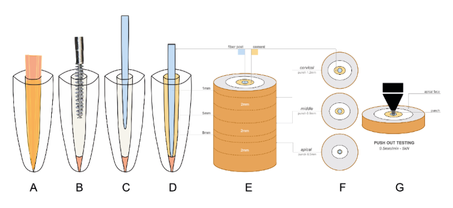
Figure 1. Illustrative diagram of the methodology.
EXPERIMENTAL GROUPS
Immediately, the gutta-percha was removed by cutting with heat, leaving 4 mm of apical sealing, using endodontic condensers. The dentin of the post space was initially cleaned with a brush (Microbrush; KG Sorensen, São Paulo, Brazil) and then with a root canal cleaning brush (MKLife, Brazil). Illustrative diagram in Figure 1.B. Different solutions were introduced for analysis (n=40): ET (95% ethanol), AA (amyl acetate), AC (99% acetone), and EX (experimental solution of amyl acetate, ethanol, and acetone in 1:1:1 proportions).
Twenty specimens from each group were evaluated for residual sealer persistence using scanning electron microscopy (SEM), and ten specimens from each group were subjected to bond strength testing of the universal adhesive system using the push-out method. The specimens were subdivided based on the type of endodontic sealer used: epoxy resin-based or bioceramic.
RESIDUAL SEALER PERSISTENCE EVALUATION
In ten roots from each group, two longitudinal grooves were made on the buccal and palatal surfaces using a low-speed diamond disc. The roots were split with a chisel, and the distal section was used for microscopic analysis. The specimens were mounted on metal stubs, gold-coated (single cycle of 120 s), and evaluated using a scanning electron microscope (EVO 10MA; Carl Zeiss, Germany) operating at 20 kV.
Four images of the root surface in the post space were taken at 500x magnification, captured by the same operator. For qualitative analysis, two blinded and calibrated examiners (k=0.93) scored the residual sealer persistence using a four-point scale (14): Score 1, absence or slight presence of sealer residues with visible dentinal tubule openings; Score 2, slight presence of residues, with more than 50% of the dentin surface free of residues; Score 3, moderate presence of residues, with <50% of the dentin surface free of residues; Score 4, intense presence of residues, with dentinal tubule openings almost or completely obstructed. Additionally, an extra image at 1500x magnification was taken from the same site to count open dentinal tubules using Image J software (version 1.52, Maryland, USA) (15).
FIBER POST CEMENTATION
The post space was standardized to a length of 11 mm with Peeso reamers #4, #5, and #6 (MKLife, Brazil) and standardized with a specific drill (Whitepost System DC 3, FGM, Brazil); followed by washing with the evaluated substances, final rinse with distilled water, drying with paper points, application of universal adhesive (Scotchbond Universal; 3M ESPE, USA) with friction for 30 seconds, solvent evaporation with compressed air for 30 seconds, removal of excess with paper cones, and light curing for 20 seconds with a LED device (Valo, Ultradent, USA). The fiber posts' surfaces (White Post DC #3, FGM) were cleaned with 70% ethanol. Silane (Prosil, FGM) was applied and dried with air for 1 minute. Then, the universal adhesive (Scotchbond Universal; 3M ESPE, USA) was applied and light-cured for 20 seconds with a LED device (Valo, Ultradent, USA). Relyx Ultimate cement (3M ESPE, USA) was inserted into the root canal with the fiber post, and light-cured for 40 seconds in various positions (buccal, lingual, mesial, distal, and occlusal) (13). Illustrative diagram in Figure 1.C and Figure 1.D.
BOND STRENGTH EVALUATION
After 24 hours, the roots were vertically positioned in a PVC matrix (21.3 mm x 20.0 mm) with evaluation on a parallelometer. The samples were sectioned perpendicularly to the longitudinal axis using a diamond disc connected to a hard tissue cutting machine (OCP 100 LC; Odeme Dental Research, Brazil). Three sections of 2.0±0.1 mm thickness were obtained from the apical, middle, and cervical thirds. Illustrative diagram in Figure 1.E.
The sections were cleaned with ultrasound and subjected to push-out bond strength testing on an electromechanical testing machine (OM150; Odeme Dental Research, Brazil) with a 5 kN load cell at 0.5 mm/min until the complete displacement of the fiber post. Punch diameters of 1.2, 0.9, and 0.5 mm were used for the cervical, middle, and apical thirds, respectively (13). Illustrative diagram in Figure 1.F and Figure 1.G.
The force required was measured in Newtons (N) and converted to bond strength (MPa) using the formula MPa=F/AD. The adhesion area (AD) was calculated as AD=π•(R+r)•g, where R is the cervical radius, r the apical radius, and g the relative height of the inverted cone.
FAILURE MODE ANALYSIS
The sections were evaluated under a stereomicroscope (Leica DFC295 connected to Leica S8 APO) at 10× magnification to determine the failure type 13. The failure mode was categorized as follows: type 1 (adhesive 1), when failure occurred between the post and the cement; type 2 (adhesive 2), between the dentin and the cement; type 3 (cohesive), within the cement; and type 4 (mixed), when both types of failure were present.
DATA ANALYSIS
Statistical analysis was performed using Jamovi software (version 2.3.18.0). Homoscedasticity was verified with the Shapiro-Wilk test. Tubule count and bond strength were analyzed using ANOVA for independent samples, followed by Tukey’s post-hoc test. Residual sealer scores were evaluated with the Kruskal-Wallis test and Dunn's post-hoc analysis, with a significance level of 5%.
RESULTS
Table 1 presents the outcomes for the epoxy resin-based cement. The protocols utilizing amyl acetate (AA) and acetone (AC) showed a significantly higher number of open dentinal tubules compared to the ethanol (ET) and experimental solution (EX) protocols, which exhibited the least tubule opening (p<0.05). Qualitative analysis revealed greater cement residue persistence in the ET and EX protocols, while the AC protocol resulted in the least amount of residue, although no statistically significant differences were observed between the cleaning protocols (p>0.05). Figure 2 provides a representative image of the dentinal substrate with epoxy resin-based cement residues according to the applied cleaning protocol.
Table 2 illustrates the results for the bioceramic cement. The ET and EX protocols achieved greater opening of the intraradicular dentinal tubules (p<0.05), whereas the AA and AC protocols demonstrated less tubule opening (p<0.05). Qualitative analysis indicated greater residue persistence with the AC protocol, although no statistically significant differences were found among the protocols (p>0.05). Figure 2 also presents an image of the dentinal substrate with bioceramic cement based on the applied cleaning protocol.
Table 3 shows that the ET protocol provided the most consistent results regarding bond strength in the apical third of the root canal when epoxy resin-based cement was used (p<0.05). However, no significant differences were found when compared to the AA and EX protocols (p>0.05). In contrast, the AC protocol exhibited the poorest bond strength results in the apical third (p<0.05). No statistically significant differences were found in the cervical and middle thirds across the cleaning protocols (p>0.05). In Figure 3, for the epoxy resin-based cement, mixed fractures predominated in the cervical third for the ET and AA protocols, while cohesive fractures in the resin were more frequent in the AC and EX protocols. In the middle third, cohesive resin fractures were dominant in the AA, AC, and EX protocols, while cohesive dentin fractures were more common in the ET protocol. In the apical third, cohesive resin fractures predominated in the ET, AA, and EX protocols.
Finally, Table 4 highlights that the ET and EX protocols yielded the best results in terms of bond strength in the cervical third when bioceramic cement was used (p<0.05). The ET protocol achieved the best results in the apical third (p<0.05), although no significant differences were observed when compared to the AA and EX protocols (p>0.05). In the middle third, no statistically significant differences were found among the evaluated protocols (p>0.05). Figure 3 shows that cohesive resin fractures predominated in the cervical thirds with the ET and AA protocols, whereas mixed fractures were more common with the AC protocol. Cohesive dentin fractures were more frequent with the EX solution. In the middle third, cohesive resin fractures predominated in the AC and EX protocols, while cohesive dentin fractures were more common in the ET protocol. In the apical third, mixed fractures were predominant across most protocols (ET, AA, and AC).
Table 1. Open dentin tubules and persistence of epoxy resin-based cement residues in intracanal dentin, according to the evaluated cleaning protocols
|
Cleaning protocols |
ET |
AA |
AC |
EX |
|
|
Open dentin tubules |
Mean |
32.80ᵇ |
49.40ᵃ |
57.60ᵃ |
24.00ᵇ |
|
SD |
3.42 |
3.58 |
3.91 |
3.61 |
|
|
CI |
30.68-34.92 |
47.18-51.62 |
55.18-60.02 |
21.77-26.23 |
|
|
Persistence of residues |
Median |
3 |
3 |
2 |
3 |
|
min-max |
3-4 |
2-3 |
2-3 |
3-4 |
|
|
1Q-3Q |
3.0-4.0 |
2.0-3.0 |
2.0-3.0 |
3.0-3.0 |
|
ᵃᵇ Different lowercase letters within the same row mean statistically significant difference (p>0.05). SD, standard deviation; CI, confidence intervals; min, minimum value; max, maximum value; 1Q y 3Q, first and third quartile. ET, ethanol; AA, amyl acetate; AC, acetone; EX, experimental solution (ethanol + amyl acetate + acetone).

Figure 2. Representative images of the dentin substrate according to endodontic sealers and cleaning protocols used. ET, ethanol; AA, amyl acetate; AC, acetone; EX, experimental solution (ethanol + amyl acetate + acetone). Scales: 20μm and 50μm.
Table 2. Open dentin tubules and persistence of bioceramic cement residues in intracanal dentin, according to the evaluated cleaning protocol.
|
Cleaning protocols |
ET |
AA |
AC |
EX |
|
|
Open dentin tubules |
Mean |
40.80ᵃ |
27.60ᵇ |
16.20ᵇ |
44.40ᵃ |
|
SD |
3.11 |
4.88 |
3.03 |
4.77 |
|
|
CI |
38.87-42.73 |
24.58-30.62 |
14.32-18.08 |
41.44-47.36 |
|
|
Persistence of residues |
Median |
3 |
3 |
4 |
3 |
|
min-max |
3-4 |
3-4 |
3-4 |
3-4 |
|
|
1Q-3Q |
3.0-4.0 |
3.0-4.0 |
4.0-4.0 |
3.0-4.0 |
|
ᵃᵇ Different lowercase letters within the same row mean statistically significant difference (p>0.05). SD, standard deviation; CI, confidence intervals; min, minimum value; max, maximum value; 1Q y 3Q, first and third quartile. ET, ethanol; AA, amyl acetate; AC, acetone; EX, experimental solution (ethanol + amyl acetate + acetone).
Table 3. Mean and standard deviation of bond strength according to the cleaning protocols for epoxy resin-based cement.
|
Protocol |
ET |
AA |
AC |
EX |
|
Cervical |
9.22 |
7.91 |
6.67 |
8.70 |
|
(0.88) |
(1.00) |
(1.35) |
(1.66) |
|
|
Middle |
5.27 |
5.47 |
2.63 |
2.67 |
|
(1.20) |
(1.31) |
(1.51) |
(1.30) |
|
|
Apical |
6.80ᵃ |
3.97ᵃᵇ |
1.37ᵇ |
4.27ᵃᵇ |
|
(1.91) |
(0.15) |
(0.43) |
(1.00) |
ᵃᵇDifferent lowercase letters within the same row mean statistically significant difference (p>0.05). ET, ethanol; AA, amyl acetate; AC, acetone; EX, experimental solution (ethanol + amyl acetate + acetone).

Figure 3. Frequency distribution of the failure modes according to the evaluated cleaning protocols. ET, ethanol; AA, amyl acetate; AC, acetone; EX, experimental solution (ethanol + amyl acetate + acetone). Type 1: adhesive, resin/dentin; Type 2: cohesive in dentin; Type 3: cohesive in resin; and Type 4: mixed.
Table 4. Mean and standard deviation of bond strength according to the cleaning protocols for bioceramic cement.
|
Protocol |
ET |
AA |
AC |
EX |
|
Cervical |
7.01ᵃ |
1.34ᵇ |
3.31ᵇ |
5.83ᵃ |
|
(1.83) |
(0.93) |
(0.97) |
(0.95) |
|
|
Middle |
3.17 |
1.06 |
3.19 |
3.44 |
|
(1.30) |
(1.28) |
(1.00) |
(1.45) |
|
|
Apical |
6.39ᵃ |
4.72ᵃᵇ |
1.73ᵇ |
3.89ᵃᵇ |
|
(1.96) |
(1.30) |
(0.68) |
(1.88) |
ᵃᵇDifferent lowercase letters within the same row mean statistically significant difference (p > 0.05). ET, ethanol; AA, amyl acetate; AC, acetone; EX, experimental solution (ethanol + amyl acetate + acetone).
DISCUSSION
Our results showed significant variations in both the persistence of residues on the intraradicular dentin substrate and the bond strength of the universal cementation system, depending on the type of endodontic cement and the cleaning protocols used. For the epoxy resin-based cement, although the protocols with ethanol and the experimental solution (ethanol, amyl acetate, and acetone) showed greater residue persistence, they achieved consistent bond strength values. In the case of bioceramic cement, these same protocols exhibited lower residue persistence and the best bond strength values. These findings allow us to reject the null hypothesis.
In the cleaning protocols applied to dentin obturated with epoxy resin-based cement, the results highlighted the superiority of amyl acetate and acetone for cleaning the dentin substrate compared to ethanol and the experimental solution. The effectiveness of amyl acetate in removing residues of epoxy resin-based cements has already been reported in the literature (8), although in studies focused on intracameral dentin. The combination of amyl acetate with acetone and ethanol has shown greater cleaning efficacy in the pulpal dentin (9), which contrasts with our results, where the experimental solution, which shared this composition, presented greater residue persistence in intraradicular dentin.
Conversely, the ethanol protocol, which demonstrated limited capacity to remove residues from the dentin substrate, as noted in the literature (10), yielded consistent results in bond strength of the universal cementation system for fiberglass posts, especially in the apical third. This outcome may be attributed to ethanol enhancing the wettability of the dentin, facilitating greater penetration of adhesive monomers, which stabilizes the hybrid layer (16,17) and more effectively seals the collagen matrix (18). Furthermore, the self-etch strategy applied to the dentin likely favored wet bonding, optimizing interaction with ethanol (19,20). Similar phenomena were observed with amyl acetate and the experimental solution, which also yielded significant bond strength values under the same conditions.
The epoxy resin residues present mainly in the protocols that included ethanol could also have influenced the bond strength results. Previous studies have demonstrated that the use of epoxy resin-based cements significantly enhances the adhesion of fiber posts (21,22), compared to eugenol-based cements and calcium silicate-based bioceramics (23). However, the acetone protocol exhibited the lowest bond strength values in radicular canals obturated with epoxy resin, likely due to acetone's interaction with the dentin substrate not providing adequate mechanical support, creating a thin adhesive layer that generated cracks at the interface (24) and negatively affecting dentin wettability (25).
Regarding bioceramic cements, the protocols with ethanol and the experimental solution were superior in cleaning the dentin substrate compared to amyl acetate and acetone. Although the effectiveness of ethanol in removing residues from bioceramic cement has not been extensively documented, some studies suggest that a combination of ethanol, amyl acetate, and acetone could improve the cleaning of these types of cements (26).
Unlike epoxy resin cements, which contain synthetic polymers with strong adhesive properties and chemical resistance (27,28), bioceramic cements are primarily composed of calcium silicate, known for its bioactivity (3). The solubility of calcium silicate is influenced by its structural characteristics and environmental conditions, such as pH and temperature (29), which explains the variability in solubility of these cements (30). Our results show that, although there is no prior evidence regarding the efficacy of ethanol or the experimental solution in removing residues from bioceramic cements, these protocols managed to eliminate the highest amount of residues. The mechanical cleaning applied in all protocols, using a low-speed activated intraradicular brush (12), may have favored this effectiveness.
Moreover, the protocols with ethanol and the experimental solution obtained the most consistent bond strength results with bioceramic cement. As mentioned, the presence of ethanol and the application of a self-etch adhesive strategy on the dentin likely enhanced wettability, facilitating the penetration of adhesive monomers and stabilizing the hybrid layer (16-20). Furthermore, the calcium silicate residues from the bioceramic cement could have promoted adhesion of the universal adhesive system (31), used in our study. Calcium silicate encourages mineral deposition, improving the interface between adhesive systems and dental substrates (32,33), in addition to providing a bioactive surface that could favorably interact with adhesive components (34).
This study was conducted on intraradicular dentin from bovine incisors, which implies limitations due to differences in organic and inorganic content compared to human dentin (35), as well as in the diameter of dentinal tubules and intertubular distance (36,37), factors that could have influenced the bond strength results. Bovine dentin was chosen due to its accessibility (38) and to avoid ethical issues associated with using human teeth (37). Furthermore, bovine dentin exhibits similar mechanical properties to human dentin (39) and provides a more uniform substrate, reducing variability in experimental results (38).
We can emphasize that cleaning protocols based on ethanol and a combination of various solutions, along with mechanical cleaning, proved effective in removing residues of bioceramic cement from intraradicular dentin, ensuring adequate bond strength in intraradicular restorative treatments with universal adhesive systems in a self-etch strategy. However, further studies are needed to evaluate the durability of the adhesive interface in endodontically treated teeth.
Finally, although ethanol was not the most effective in removing residues from epoxy resin-based cements, it demonstrated consistency in cleaning bioceramic cements and achieved adequate bond strength with fiberglass posts, regardless of the type of endodontic cement. The experimental solution also showed great potential as a cleaning agent, warranting further studies to confirm its effectiveness.
CONCLUSION
This study demonstrates that chemical-mechanical cleaning protocols using ethanol and an experimental solution based on ethanol, amyl acetate, and acetone are effective in removing residues of bioceramic cements from intraradicular dentin, achieving adequate bond strength of the universal adhesive system for fiberglass posts. Although a greater persistence of residues was observed with epoxy resin-based cements, the evaluated protocols also ensured satisfactory bond strength in these cases.
CONFLICT OF INTEREST: No conflict of interest.
FUNDING: This work was supported by the Jorge Basadre Grohmann National University (UNJBG), Peru, under Finance Code 13818-2024.
AUTHOR CONTRIBUTION STATEMENT: Conceptualization and design: J.B.T., G.M.C.N. and M.C.K.; Literature review: I.D.M.C., E.C.Z.M. and W.G.E.O.; Methodology and validation: I.D.M.C., E.C.Z.M. and W.G.E.O.; Formal analysis: J.B.T., W.G.E.O., M.C.K. and G.M.C.N.; Investigation and data collection: I.D.M.C. and E.C.Z.M.; Resources: J.B.T., W.G.E.O. and G.M.C.N.; Data analysis and interpretation: W.G.E.O., M.C.K. and G.M.C.N.; Writing-review and editing: J.B.T., W.G.E.O., M.C.K. and G.M.C.N.; Supervision: W.G.E.O. and M.C.K.; Project administration: J.B.T. and G.M.C.N.
ACKNOWLEDGMENTS: The authors would like to thank the Jorge Basadre Grohmann National University (UNJBG) and the Vice-Rectorate for Research of the Catholic University of Santa María (UCSM) for their support in the development of this study.
REFERENCES
1. De Moor R.J., De Boever J.G. The sealing ability of an epoxy resin root canal sealer used with five gutta-percha obturation techniques. Endod Dent Traumatol [Internet]. 2000 Dec; 16 (6): 291-7. Available from: http://dx.doi.org/10.1034/j.1600-9657.2000.016006291.x
2. Troiano G., Perrone D., Dioguardi M., Buonavoglia A., Ardito F., Lo Muzio L. In vitro evaluation of the cytotoxic activity of three epoxy resin-based endodontic sealers. Dent Mater J [Internet]. 2018 Jun 8; 37 (3): 374-8. Available from: http://dx.doi.org/10.4012/dmj.2017-148
3. Abreu Betinelli G.A. de, Modolon H.B., Wermuth T.B., Raupp-Pereira F., Klegues Montedo O.R., Vassen A.B., et al. Combustion synthesis of nanostructured calcium silicates: A new approach to develop bioceramic cements in endodontics. Ceram Int [Internet]. 2024 Feb; 50 (3): 4544-52. Available from: https://linkinghub.elsevier.com/retrieve/pii/S0272884223036490
4. Sheela S., Nassar M., AlGhalban F.M., Gorduysus M.O. In Vitro Cytotoxicity and Mineralization Potential of an Endodontic Bioceramic Material. Eur J Dent [Internet]. 2023 May; 17 (2): 548-55. Available from: http://dx.doi.org/10.1055/s-0042-1750778
5. Sanz J.L., López-García S., Rodríguez-Lozano F.J., Melo M., Lozano A., Llena C., et al. Cytocompatibility and bioactive potential of AH Plus Bioceramic Sealer: An in vitro study. Int Endod J [Internet]. 2022 Oct; 55 (10): 1066-80. Available from: http://dx.doi.org/10.1111/iej.13805
6. Olcay K., Taşli P.N., Güven E.P., Ülker G.M.Y., Öğüt E.E., Çiftçioğlu E., et al. Effect of a novel bioceramic root canal sealer on the angiogenesis-enhancing potential of assorted human odontogenic stem cells compared with principal tricalcium silicate-based cements. J Appl Oral Sci [Internet]. 2020 Jan 10; 28: e20190215. Available from: http://dx.doi.org/10.1590/1678-7757-2019-0215
7. Devroey S., Calberson F., Meire M. The efficacy of different cleaning protocols for the sealer-contaminated access cavity. Clin Oral Investig [Internet]. 2020 Nov; 24 (11): 4101-7. Available from: https://link.springer.com/10.1007/s00784-020-03283-8
8. Zaniboni J.F., de Souza V., Escalante-Otárola W.G., Porto T.S., Godoy E.F., Kuga M.C. Impact of cleansing protocols to remove endodontic sealer residues on the adhesive interface: Bonding with universal adhesive systems. J Esthet Restor Dent [Internet]. 2022 Oct; 34 (7): 1077-84. Available from: http://dx.doi.org/10.1111/jerd.12924
9. Zaniboni J.F., de Souza V., Escalante-Otárola W.G., Leandrin T.P., Fernández Godoy E., Besegato J.F., et al. Cleaning and microstructural effects of amyl acetate on pulp chamber dentin impregnated with epoxy resin-based endodontic sealer. J Esthet Restor Dent [Internet]. 2022 Dec; 34 (8): 1282-9. Available from: http://dx.doi.org/10.1111/jerd.12966
10. Kuga M.C., Só M.V.R., De Faria-júnior N.B., Keine K.C., Faria G., Fabricio S., et al. Persistence of resinous cement residues in dentin treated with different chemical removal protocols. Microsc Res Tech [Internet]. 2012 Jul; 75 (7): 982-5. Available from: https://analyticalsciencejournals.onlinelibrary.wiley.com/doi/10.1002/jemt.22023
11. Alencar C.M., Verbicário Dos Santos J., Jassé F., Dos Santos G.O., Escalante-Otárola W.G., Castro-Nuñez G.M., et al. Protocols for Mechanical Cleaning of the Post Space on the Bond Strength Between Root Dentin and Cementation System. Oper Dent [Internet]. 2021 Jul 1; 46 (4): 467-75. Available from: http://dx.doi.org/10.2341/20-194-L
12. Castro-Núnez G.M., Dos Santos J.R.E.V., Zaniboni J.F., Escalante-Otárola W.G., Porto T.S., Kuga M.C. Effect of mechanical cleaning protocols in the fiber post space on the adhesive interface between universal adhesive and root dentin. Microsc Res Tech [Internet]. 2022 Jun; 85 (6): 2131-9. Available from: https://analyticalsciencejournals.onlinelibrary.wiley.com/doi/10.1002/jemt.24071
13. Lima R.O., Barros A.P.O., Alencar C.D.M., Pereira K.D.F., Galvani L.D., Vaz L.G., et al. Influence of using different translucent composite resins for customizing fiber post on the bond strength of self-adhesive cement to root dentin. Dent Mater J [Internet]. 2024 Jan 30; 43 (1): 112-8. Available from: http://dx.doi.org/10.4012/dmj.2023-184
14. Vitória M.S., de Almeida E.N.M., Barros A.P.O., de Oliveira E.C.G., Costa J.L. de S.G., Dantas A.A.R., et al. Evaluation of different mechanical cleaning protocols associated with 2.5% sodium hypochlorite in the removal of residues from the post space. J Conserv Dent Endod [Internet]. 2024 Mar; 27 (3): 274-9. Available from: http://dx.doi.org/10.4103/JCDE.JCDE_324_23
15. Escalante-Otárola W.G., Castro-Núñez G.M., Leandrim T.P., Alencar C.M., de Albuquerque Jassé F.F., Kuga M.C. Effects of Remineralizing Agents Based on Calcium Phosphate, Sodium Phosphate, or Sodium Fluoride on Eroded Cervical Dentin. Oper Dent [Internet]. 2021 Nov 1; 46 (6): E296-306. Available from: http://dx.doi.org/10.2341/20-209-L
16. Li F., Liu X.Y., Zhang L., Kang J.J., Chen J.H. Ethanol-wet bonding technique may enhance the bonding performance of contemporary etch-and-rinse dental adhesives. J Adhes Dent [Internet]. 2012 Apr; 14 (2): 113-20. Available from: http://dx.doi.org/10.3290/j.jad.a21853
17. Ayar M.K. A review of ethanol wet-bonding: Principles and techniques. Eur J Dent [Internet]. 2016 Jan-Mar; 10 (1): 155-9. Available from: http://dx.doi.org/10.4103/1305-7456.175687
18. Bitter K., Aschendorff L., Neumann K., Blunck U., Sterzenbach G. Do chlorhexidine and ethanol improve bond strength and durability of adhesion of fiber posts inside the root canal? Clin Oral Investig [Internet]. 2014 Apr; 18 (3): 927-34. Available from: http://dx.doi.org/10.1007/s00784-013-1040-1
19. Talungchit S., Jessop J.L.P., Cobb D.S., Qian F., Geraldeli S., Pashley D.H., et al. Ethanol-wet bonding and chlorhexidine improve resin-dentin bond durability: quantitative analysis using raman spectroscopy. J Adhes Dent [Internet]. 2014 Oct; 16 (5): 441-50. Available from: http://dx.doi.org/10.3290/j.jad.a32695
20. Hosaka K., Nishitani Y., Tagami J., Yoshiyama M., Brackett W.W., Agee K.A., et al. Durability of resin-dentin bonds to water- vs. ethanol-saturated dentin. J Dent Res [Internet]. 2009 Feb; 88 (2): 146-51. Available from: http://dx.doi.org/10.1177/0022034508328910
21. Alqarni H., Khan S., Alzaid A.A., Alfaifi M., Alsayed H., Alshahrani N., et al. Effect of silicone-based sealers on fiber post retention: An in vitro study. J Prosthodont [Internet]. 2023 Nov 15; Available from: http://dx.doi.org/10.1111/jopr.13799
22. Prado M., Marques J.N., Pereira G.D., da Silva E.M., Simão R.A. Evaluation of different surface treatments on fiber post cemented with a self-adhesive system. Mater Sci Eng C Mater Biol Appl [Internet]. 2017 Aug 1; 77: 257-62. Available from: http://dx.doi.org/10.1016/j.msec.2017.03.141
23. Vilas-Boas D.A., Grazziotin-Soares R., Ardenghi D.M., Bauer J., de Souza P.O., de Miranda Candeiro G.T., et al. Effect of different endodontic sealers and time of cementation on push-out bond strength of fiber posts. Clin Oral Investig [Internet]. 2018 Apr; 22 (3): 1403-9. Available from: http://dx.doi.org/10.1007/s00784-017-2230-z
24. Cho B.H., Dickens S.H. Effects of the acetone content of single solution dentin bonding agents on the adhesive layer thickness and the microtensile bond strength. Dent Mater [Internet]. 2004 Feb; 20 (2): 107-15. Available from: http://dx.doi.org/10.1016/s0109-5641(03)00071-x
25. Soares J., Perdigão J., Chrispim B., Lopes G.C. Effect of Extra Hydrophobic Resin Layer on Bonding of Universal Adhesive Systems to Enamel. Oper Dent [Internet]. 2023 Mar 1; 48 (2): E48-59. Available from: http://dx.doi.org/10.2341/21-140-L
26. Oliveira Barros A.P., Freitas A.P.A.R.A., Kokol F.G.O., de Souza E.M.P., Junior A.J., de Melo Alencar C., et al. Influence of the use of a mixed solution of equal amounts of amyl acetate, acetone, and ethanol on the cleaning of endodontic sealer residues on the bond strength of the fiber post cementation system: A laboratory investigation. Open Dent J [Internet]. 2024 Mar 5; 18 (1). Available from: https://opendentistryjournal.com/VOLUME/18/ELOCATOR/e18742106279970/
27. Elias V.V., Lima R.B., Lucisano M.P., Araujo L.D.C., Pucinelli C.M., Nelson-Filho P., et al. Inflammatory response to bioceramic and epoxy resin-based endodontic sealers implanted in mice subcutaneous tissue: An in vivo study. Microsc Res Tech [Internet]. 2024 Oct; 87 (10): 2447-58. Available from: http://dx.doi.org/10.1002/jemt.24631
28. Kwak S.W., Koo J., Song M., Jang I.H., Gambarini G., Kim H.C. Physicochemical Properties and Biocompatibility of Various Bioceramic Root Canal Sealers: In Vitro Study. J Endod [Internet]. 2023 Jul; 49 (7): 871-9. Available from: http://dx.doi.org/10.1016/j.joen.2023.05.013
29. Chen J.J., Thomas J.J., Taylor H.F.W., Jennings H.M. Solubility and structure of calcium silicate hydrate. Cem Concr Res [Internet]. 2004 Sep; 34 (9): 1499-519. Available from: https://linkinghub.elsevier.com/retrieve/pii/S000888460400211X
30. Torres F.F.E., Guerreiro-Tanomaru J.M., Bosso-Martelo R., Chavez-Andrade G.M., Tanomaru Filho M. Solubility, porosity and fluid uptake of calcium silicate-based cements. J Appl Oral Sci [Internet]. 2018 May 21; 26: e20170465. Available from: http://dx.doi.org/10.1590/1678-7757-2017-0465
31. Naiboğlu P., Koşar T., Yücel A.Ç. Shear bond strength of calcium silicate-based cements to composite resin using a universal adhesive in different application modes: an in vitro study. Aust Dent J [Internet]. 2024 Jun; 69 (2): 102-11. Available from: http://dx.doi.org/10.1111/adj.12990
32. Balbinot G.S., Leitune V.C.B., Ogliari F.A., Collares F.M. Niobium silicate particles promote in vitro mineral deposition on dental adhesive resins. J Dent [Internet]. 2020 Oct; 101: 103449. Available from: http://dx.doi.org/10.1016/j.jdent.2020.103449
33. Pires-de-Souza F. de C.P., Tonani-Torrieri R., Geng Vivanco R., Arruda C.N.F. de, Geraldeli S., Sinhoreti M.A.C., et al. Effect of Incorporation of Bioactive Glass-Ceramic into Self-etch Adhesives. J Adhes Dent [Internet]. 2022 Apr 13; 24: 195-202. Available from: http://dx.doi.org/10.3290/j.jad.b2916451
34. Aggarwal V., Bhasin S.S. Application of Calcium Silicate Materials After Acid Etching May Preserve Resin-Dentin Bonds. Oper Dent [Internet]. 2018 Jun 28; 43 (5): E243-52. Available from: http://dx.doi.org/10.2341/17-306-L
35. Enrich-Essvein T., Benavides-Reyes C., Álvarez-Lloret P., Bolaños-Carmona M.V., Rodríguez-Navarro A.B., González-López S. Influence of de-remineralization process on chemical, microstructural, and mechanical properties of human and bovine dentin. Clin Oral Investig [Internet]. 2021 Mar; 25 (3): 841-9. Available from: http://dx.doi.org/10.1007/s00784-020-03371-9
36. Dehghani Nazhvani A., Dehghanpour Frashah H., Haddadi P., Dehghani Nazhvani F. Ultrastructural and Chemical Composition of Dentin and Enamel in Lab Animals. J Dent [Internet]. 2019 Sep; 20 (3): 178-83. Available from: http://dx.doi.org/10.30476/DENTJODS.2019.44912
37. Wegehaupt F., Gries D., Wiegand A., Attin T. Is bovine dentine an appropriate substitute for human dentine in erosion/abrasion tests? J Oral Rehabil [Internet]. 2008 May; 35 (5): 390-4. Available from: http://dx.doi.org/10.1111/j.1365-2842.2007.01843.x
38. Silva E.J.N.L., Carvalho N.K., Prado M.C., Senna P.M., Souza E.M., De-Deus G. Bovine teeth can reliably substitute human dentine in an intra-tooth push-out bond strength model? Int Endod J [Internet]. 2019 Jul; 52 (7): 1063-9. Available from: http://dx.doi.org/10.1111/iej.13085
39. Inoue T., Miyazaki T., Nishimura F. Tensile strength and durability of bovine dentin. Dent Mater J [Internet]. 2007 May; 26 (3): 348-54. Available from: http://dx.doi.org/10.4012/dmj.26.348
 Odovtos -Int J Dent Sc endoses to CC-BY-NC-SA 4.0.