

Keywords
AH-Plus; BioRoot RCS; Ligamento periodontal; Sellador endodóntico; Obturación endodóntica; Citotoxicidad; Biocompatibilidad.
How to Cite
Abstract
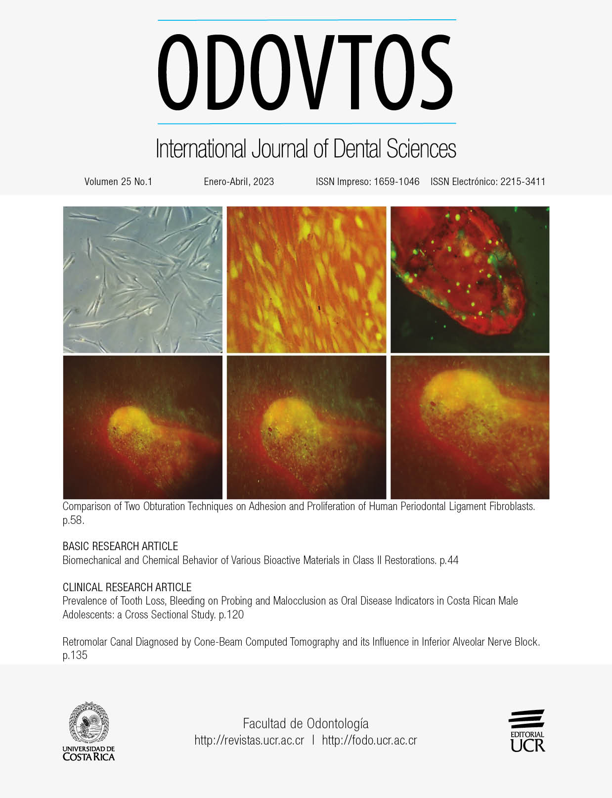
The present study aimed to compare the adhesion and proliferation of human periodontal ligament fibroblasts (hPDL) in transverse sections of the teeth sealed with two different obturation techniques, BioRoot RCS/hydraulic obturation (HO) and AH-Plus/continuous-wave condensation (CWC). The techniques were tested using an in vitro model to simulate the interaction between periodontal tissues and the materials. The root canals were instrumented and sterilized. A total of 15 samples were obturated with BioRoot RCS/HO and 15 samples with AH-Plus/CWC. Then, roots were sectioned to obtain obturated teeth slices, and hPDL cells were seeded onto the root slices. The results were obtained at intervals of 4 and 24 h for cell adhesion; and at 3,7,14, and 21 days for cell proliferation. Empty cell culture plates were use as controls. The cell adhesion was increased at 4 and 24 h for both groups, with an increased response observed in the BioRoot RCS/HO group (p<0.05). The difference in cell proliferation was also found between experimental groups. After 14 days of culture, BioRoot RCS/HO group showed an increase response than control and AH-Plus/CWC groups (p<0.05), and after 21 days both groups behaved better than control group, with an increased response observed in the BioRoot RCS/HO group. This study demonstrated that both root canal sealers allow the attach and growth of periodontal ligament fibroblasts, with an increased biological response in the BioRoot RCS/HO group.
References
Jeanneau C., Giraud T., Laurent P., About I. Bioroot RCS extracts modulate the early mechanisms of periodontal inflammation and regeneration. J Endod 2019; 45: 1016-1023.
López-García S., Pecci-Lloret M.R., Guerrero-Gironés J. et al. Comparative cytocompatibility and mineralization potential of Bio-C sealer and Total Fill BC sealer. Materials (Basel) 2019; 12 (19): 3087.
Whitworth J. Methods of filling root canals: principles and practices. Endod Topics 2005; 12: 2-24.
Ricucci D., Rôcas I.N., Alves F., Loghin S., Siqueira J.F. Apically extruded sealers: Fate and influence on treatment outcome. J Endod 2016; 42: 243-249.
Holland R., Gomes J.E. Filho, Cintra L.T.A., Queiroz Í.O.A., Estrela C. Factors affecting the periapical healing process of endodontically treated teeth. J Appl Oral Sci 2017; 25: 465-476.
Fonseca D.A., Paula A.B., Marto C.M., Coelho A., Paulo S., Martinho J.P., Carrilho E., Ferreira M.M. Biocompatibility of Root Canal Sealers: A Systematic Review of In Vitro and In Vivo Studies. aterials (Basel) 2019; 12: 4113
Lee J.K., Kwak S.W., Ha J.H., Lee W., Kim H.C. Physicochemical properties of epoxy resin-based and bioceramic-based root canal sealers. Bioinorg Chem Appl 2017; 2017: 2582849.
Huang T.H., Yang J.J., Li H., Kao C.T. The biocompatibility evaluation of epoxy resin-based root canal sealers in vitro. Biomaterials 2002; 23: 77-83.
Eldeniz A.U., Mustafa K., Ørstavik D., Dahl J.E. Cytotoxicity of new resin-, calcium hydroxide- and silicone-based root canal sealers on fibroblasts derived from human gingiva and L929 cell lines. Int Endod J 2007; 40: 329-337.
Huang F.M., Chou M.Y., Chang Y.C. Induction of cyclooxygenase-2 mRNA and protein expression by epoxy resin and zinc oxide-eugenol based root canal sealers in human osteoblastic cells. Biomaterials 2003; 24: 1869-1875.
Drukteinis S. Hydraulic calcium silicate‑based materials for root canal obturation. Int J Dent 2022; 9: 6958135
Camps J., Jeanneau C., El Ayachi I., Laurent P., About I. Bioactivity of a Calcium Silicate-based Endodontic Cement (BioRoot RCS): Interactions with Human Periodontal Ligament Cells In Vitro. J Endod 2015; 41: 1469-1473.
Accardo C., Himel V.T., Lallier T.E. A novel Gutta Flow sealer supports cell survival and attachment. J Endod 2014; 40: 231-234.
Hench L.L. The story of Bioglass. J Mater Sci Mater Med 2006; 17: 967-978.
Malhotra S., Hedge M., Shetty C. Bioceramic technology in endodontics. Br J Med Med Res 2014; 4: 2446-2454.
About I., Bottero M.J., Denato P., Camps J., Franquin J.C., Mitsiadis T.A. Human dentin production in vitro. Exp Cell Res 2000; 258: 33-41.
Feoktistova M., Geserick P., Leverkus M. Crystal violet assay for determining viability of cultured cells. Cold Spring Harb Protoc 2016; 166: 163-171.
Ishiyama M., Tominaga H., Shiga M., Sasamoto K., Ohkura Y., Ueno K. A combined assay of cell viability and in vitro cytotoxicity with a highly water-soluble tetrazolium salt, neutral red and crystal violet. Biol Pharm Bull 1996; 19:1518-1520.
Elliott J.T., Tona A., Plant A.L. Comparison of reagents for shape analysis of fixed cells by automated fluorescence microscopy. Cytometry 2003; 52A: 90-100.
Accardo C., Himel V.T., Lallier T.E. A novel GuttaFlow sealer supports cell survival and attachment. J Endod 2014; 40: 231-234.
Diomede F., Caputi S., Merciaro I., et al. Pro-inflammatory cytokine release and cell growth inhibition in primary human oral cells after exposure to endodontic sealer. Int Endod J 2014; 47: 864-872.
Silva L.A., Barnett F., Pumarola-Suñé J., Cañadas P.S., Nelson-Filho P., Silva R.A. Sealapex Xpress and Real Seal XT feature tissue compatibility in vivo. J Endod 2014; 40: 1424-1428.
Prüllage R.K., Urban K., Schäfer E., Dammaschke T. Material properties of a Tricalcium Silicate-containing, a Mineral Trioxide Aggregate-containing, and an Epoxy Resin-based Root Canal Sealer. J Endod 2016; 42: 1784-1788.
Vivan R.R., Zapata R.O., Zeferino M.A., et al. Evaluation of the physical and chemical properties of two commercial and three experimental root-end filling material. Oral Surg Oral Med Oral Pathol Oral Radiol Endod 2010; 110: 250-256.
Collado-González M., García-Bernal D., Oñate-Sánchez R.E., et al. Biocompatibility of three new calcium silicate-based endodontic sealers on human periodontal ligament stem cells. Int Endod J 2017; 50: 875-884.
Schäfer E., Köster M., Bürklein S. Percentage of gutta-percha-filled areas in canals instrumented with nickel-titanium systems and obturated with matching single cones. J Endod 2013; 39: 924-928.
Ahmed H., Ratnayake J., Cathro P., Chandler N. The effect of an additional application of sealer prior to backfilling in the Continuous Wave of Condensation technique. Aust Endod J 2022; In press.
Li G.H., Niu L.N., Selem L.C., et al. Quality of obturation achieved by an endodontic core-carrier system with crosslinked gutta-percha carrier in single-rooted canals. J Dent 2014; 42: 1124-1134.
Donnermeyer D., Ibing M., Bürklein S., Weber I., Reitze M.P., Schäfer E. Physico-Chemical Investigation of Endodontic Sealers Exposed to Simulated Intracanal Heat Application: Hydraulic Calcium Silicate-Based Sealers. Materials (Basel) 2021; 14: 728.
Chavarria-Bolanos D., Komabayashi T., Shen I., Vega-Baudrit J., Gandolfi M.G., Prati C., Montero-Aguilar M. Effects of heat on seven endodontic sealers. J Oral Sci 2022; 64: 33-39.
Urban K., Neuhaus J., Donnermeyer D., Schäfer E., Dammaschke. Solubility and pH value of 3 different root canal sealers: a long-term investigation. J Endod 2018; 44: 1736-1740.
Somma F., Cretella G., Carotenuto M., et al. Quality of thermoplasticized and single point root fillings assessed by micro-computed tomography. Int Endod J 2011; 44: 362-369.
##plugins.facebook.comentarios##

This work is licensed under a Creative Commons Attribution-NonCommercial-ShareAlike 4.0 International License.
Copyright (c) 2022 CC-BY-NC-SA 4.0