

Palabras clave
AH-Plus; BioRoot RCS; Ligamento periodontal; Sellador endodóntico; Obturación endodóntica; Citotoxicidad; Biocompatibilidad.
Cómo citar
Resumen
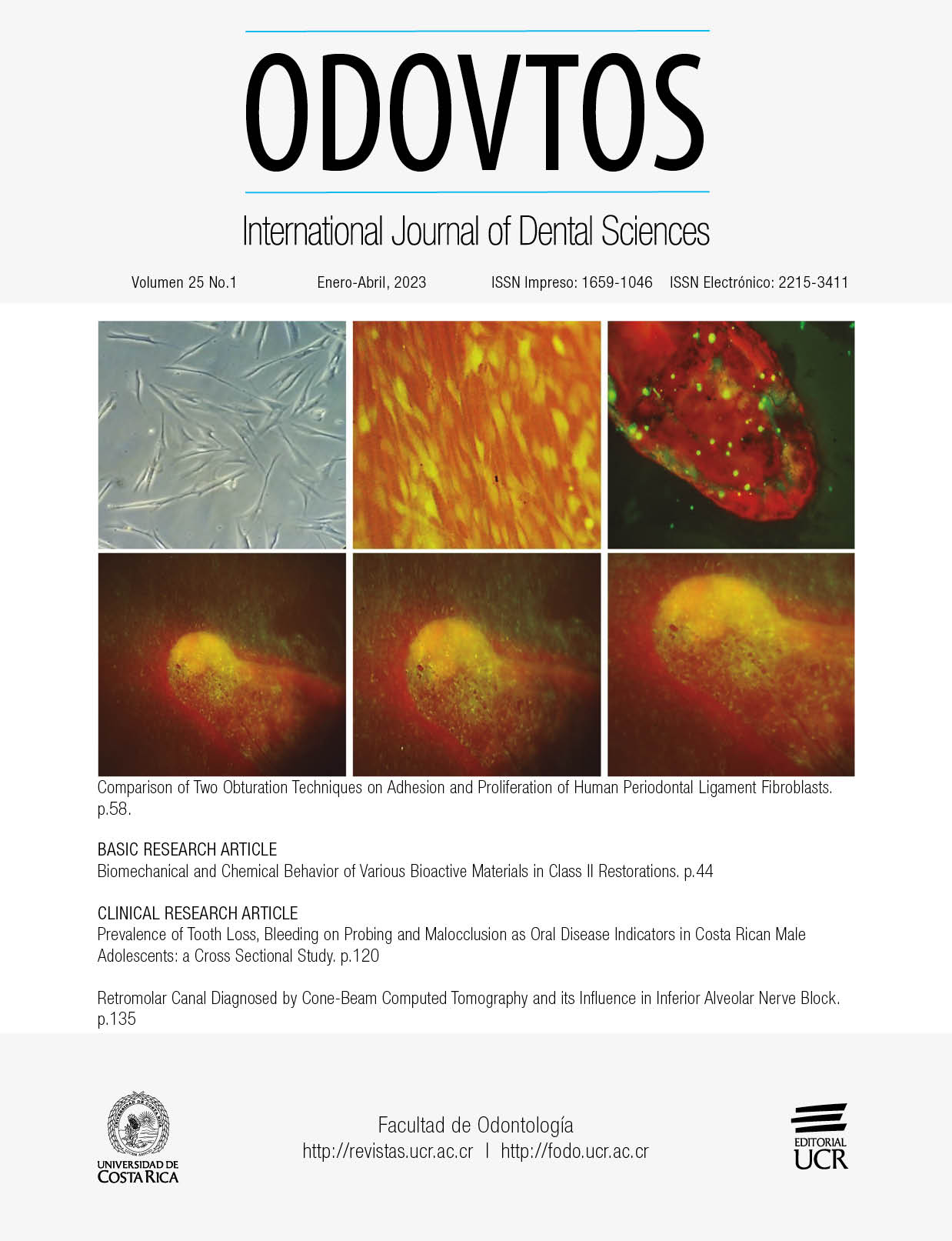
El presente estudio se enfocó en comparar la adhesión y proliferación de fibroblastos de ligamento periodontal humano (hPDL) en secciones transversales de raíces previamente obturadas con dos técnicas de obturación diferentes: obturación hidráulica empleando cono único de gutapercha y BioRoot RCS como sellador (HO), y obturación de condensación de onda continua y AH-Plus como sellador (CWC). Los selladores se usaron en un modelo in vitro que simula la interacción entre los tejidos periodontales y los materiales de obturación. Los conductos radiculares fueron instrumentados, esterilizados y obturados. La muestra se compuso de un total de 15 raíces con la técnica BioRoot RCS/HO y 15 raíces con la técnica AH-Plus/CWC. Las células de hPDL fueron sembradas en condiciones estándar de cultivo sobre las raíces seccionadas. Los resultados fueron obtenidos a intervalos de 4 y 24h para adhesión celular, y a los 3,5,7,14 y 21 días de cultivo para proliferación celular. La adhesión celular a las 4 y 24 horas mostró ser diferente para ambas técnicas en comparación con el grupo control, siendo más importante en el grupo BioRoot RCS/HO. La diferencia en la proliferación entre grupos se observó a los 14 días de cultivo, únicamente para el grupo BioRoot RCS/HO; Sin embargo para el día 21 ambas técnicas mostraron mayor proliferación celular que el grupo control, con mejor respuesta para el grupo BioRoot RCS/HO. Este estudio ha demostrado que ambos selladores de conductos permiten la adhesión y crecimiento de fibroblastos de ligamento periodontal, siendo el grupo BioRoot RCS/HO el que mostró mayor biocompatibilidad.
Citas
Jeanneau C., Giraud T., Laurent P., About I. Bioroot RCS extracts modulate the early mechanisms of periodontal inflammation and regeneration. J Endod 2019; 45: 1016-1023.
López-García S., Pecci-Lloret M.R., Guerrero-Gironés J. et al. Comparative cytocompatibility and mineralization potential of Bio-C sealer and Total Fill BC sealer. Materials (Basel) 2019; 12 (19): 3087.
Whitworth J. Methods of filling root canals: principles and practices. Endod Topics 2005; 12: 2-24.
Ricucci D., Rôcas I.N., Alves F., Loghin S., Siqueira J.F. Apically extruded sealers: Fate and influence on treatment outcome. J Endod 2016; 42: 243-249.
Holland R., Gomes J.E. Filho, Cintra L.T.A., Queiroz Í.O.A., Estrela C. Factors affecting the periapical healing process of endodontically treated teeth. J Appl Oral Sci 2017; 25: 465-476.
Fonseca D.A., Paula A.B., Marto C.M., Coelho A., Paulo S., Martinho J.P., Carrilho E., Ferreira M.M. Biocompatibility of Root Canal Sealers: A Systematic Review of In Vitro and In Vivo Studies. aterials (Basel) 2019; 12: 4113
Lee J.K., Kwak S.W., Ha J.H., Lee W., Kim H.C. Physicochemical properties of epoxy resin-based and bioceramic-based root canal sealers. Bioinorg Chem Appl 2017; 2017: 2582849.
Huang T.H., Yang J.J., Li H., Kao C.T. The biocompatibility evaluation of epoxy resin-based root canal sealers in vitro. Biomaterials 2002; 23: 77-83.
Eldeniz A.U., Mustafa K., Ørstavik D., Dahl J.E. Cytotoxicity of new resin-, calcium hydroxide- and silicone-based root canal sealers on fibroblasts derived from human gingiva and L929 cell lines. Int Endod J 2007; 40: 329-337.
Huang F.M., Chou M.Y., Chang Y.C. Induction of cyclooxygenase-2 mRNA and protein expression by epoxy resin and zinc oxide-eugenol based root canal sealers in human osteoblastic cells. Biomaterials 2003; 24: 1869-1875.
Drukteinis S. Hydraulic calcium silicate‑based materials for root canal obturation. Int J Dent 2022; 9: 6958135
Camps J., Jeanneau C., El Ayachi I., Laurent P., About I. Bioactivity of a Calcium Silicate-based Endodontic Cement (BioRoot RCS): Interactions with Human Periodontal Ligament Cells In Vitro. J Endod 2015; 41: 1469-1473.
Accardo C., Himel V.T., Lallier T.E. A novel Gutta Flow sealer supports cell survival and attachment. J Endod 2014; 40: 231-234.
Hench L.L. The story of Bioglass. J Mater Sci Mater Med 2006; 17: 967-978.
Malhotra S., Hedge M., Shetty C. Bioceramic technology in endodontics. Br J Med Med Res 2014; 4: 2446-2454.
About I., Bottero M.J., Denato P., Camps J., Franquin J.C., Mitsiadis T.A. Human dentin production in vitro. Exp Cell Res 2000; 258: 33-41.
Feoktistova M., Geserick P., Leverkus M. Crystal violet assay for determining viability of cultured cells. Cold Spring Harb Protoc 2016; 166: 163-171.
Ishiyama M., Tominaga H., Shiga M., Sasamoto K., Ohkura Y., Ueno K. A combined assay of cell viability and in vitro cytotoxicity with a highly water-soluble tetrazolium salt, neutral red and crystal violet. Biol Pharm Bull 1996; 19:1518-1520.
Elliott J.T., Tona A., Plant A.L. Comparison of reagents for shape analysis of fixed cells by automated fluorescence microscopy. Cytometry 2003; 52A: 90-100.
Accardo C., Himel V.T., Lallier T.E. A novel GuttaFlow sealer supports cell survival and attachment. J Endod 2014; 40: 231-234.
Diomede F., Caputi S., Merciaro I., et al. Pro-inflammatory cytokine release and cell growth inhibition in primary human oral cells after exposure to endodontic sealer. Int Endod J 2014; 47: 864-872.
Silva L.A., Barnett F., Pumarola-Suñé J., Cañadas P.S., Nelson-Filho P., Silva R.A. Sealapex Xpress and Real Seal XT feature tissue compatibility in vivo. J Endod 2014; 40: 1424-1428.
Prüllage R.K., Urban K., Schäfer E., Dammaschke T. Material properties of a Tricalcium Silicate-containing, a Mineral Trioxide Aggregate-containing, and an Epoxy Resin-based Root Canal Sealer. J Endod 2016; 42: 1784-1788.
Vivan R.R., Zapata R.O., Zeferino M.A., et al. Evaluation of the physical and chemical properties of two commercial and three experimental root-end filling material. Oral Surg Oral Med Oral Pathol Oral Radiol Endod 2010; 110: 250-256.
Collado-González M., García-Bernal D., Oñate-Sánchez R.E., et al. Biocompatibility of three new calcium silicate-based endodontic sealers on human periodontal ligament stem cells. Int Endod J 2017; 50: 875-884.
Schäfer E., Köster M., Bürklein S. Percentage of gutta-percha-filled areas in canals instrumented with nickel-titanium systems and obturated with matching single cones. J Endod 2013; 39: 924-928.
Ahmed H., Ratnayake J., Cathro P., Chandler N. The effect of an additional application of sealer prior to backfilling in the Continuous Wave of Condensation technique. Aust Endod J 2022; In press.
Li G.H., Niu L.N., Selem L.C., et al. Quality of obturation achieved by an endodontic core-carrier system with crosslinked gutta-percha carrier in single-rooted canals. J Dent 2014; 42: 1124-1134.
Donnermeyer D., Ibing M., Bürklein S., Weber I., Reitze M.P., Schäfer E. Physico-Chemical Investigation of Endodontic Sealers Exposed to Simulated Intracanal Heat Application: Hydraulic Calcium Silicate-Based Sealers. Materials (Basel) 2021; 14: 728.
Chavarria-Bolanos D., Komabayashi T., Shen I., Vega-Baudrit J., Gandolfi M.G., Prati C., Montero-Aguilar M. Effects of heat on seven endodontic sealers. J Oral Sci 2022; 64: 33-39.
Urban K., Neuhaus J., Donnermeyer D., Schäfer E., Dammaschke. Solubility and pH value of 3 different root canal sealers: a long-term investigation. J Endod 2018; 44: 1736-1740.
Somma F., Cretella G., Carotenuto M., et al. Quality of thermoplasticized and single point root fillings assessed by micro-computed tomography. Int Endod J 2011; 44: 362-369.
Comentarios

Esta obra está bajo una licencia internacional Creative Commons Atribución-NoComercial-CompartirIgual 4.0.
Derechos de autor 2022 CC-BY-NC-SA 4.0