Resumen
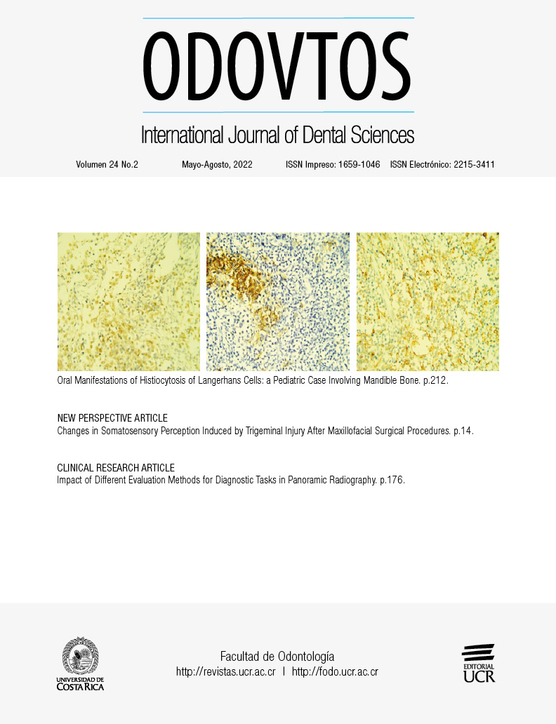
La histiocitosis de las células de Langerhans es una enfermedad poco frecuente que se caracteriza por la proliferación monoclonal y la migración de células dendríticas especiales en una variedad de órganos; lo más común es que aparezca un granuloma eosinofílico localizado, a menudo solitario, así como lesiones óseas que se producen predominantemente en pacientes pediátricos. Aunque es más frecuente en los niños menores de 15 años, este trastorno se presenta en todas las edades y se produce a una tasa de 2 a 5 casos por millón al año. La HCL es una entidad compleja; las manifestaciones clínicas pueden imitar otras afecciones comunes y, por lo tanto, se indica una evaluación exhaustiva. Dado que las manifestaciones orales son frecuentes, el diagnóstico precoz de esta patología podría ser detectado por los profesionales de la odontología. El objetivo de este reporte de caso es describir un caso de HCL que inicialmente fue mal diagnosticado y tratado por una infección dental. Esta enfermedad requiere un diagnóstico histopatológico preciso y un tratamiento oportuno; por lo tanto, es necesario sensibilizar a los dentistas para evitar un diagnóstico erróneo de las manifestaciones orales de la HCL.
Citas
Margo C.E., Goldman D.R. Langerhans cell histiocytosis. Surv Ophthalmol. 2008; 53: 332-58. doi:10.1016/j.survophthal.2008.04.007
Satter E.K., High W.A. Langerhans cell histiocytosis: a review of the current recommendations of the Histiocyte Society. Pediatr Dermatol. 2008; 25: 291-5. doi:10.1111/j.1525-1470.2008.00669.x
Allen C.E., Merad M., McClain K.L. Langerhans-cell histiocytosis. N Eng J Med. 2018, 379 (9), 856-868. doi: 10.1056/NEJMra1607548
Krooks J., Minkov M., Weatherall A.G. Langerhans cell histiocytosis in children: History, classification, pathobiology, clinical manifestations, and prognosis. J Am Acad Dermatol. 2018 Jun; 78 (6): 1035-1044. doi: 10.1016/j.jaad.2017.05.059. PMID: 29754885
Guyot-Goubin A., Donadieu J., Barkaoui M., Bellec S., Thomas C., Clavel J. Descriptive epidemiology of childhood Langerhans cell histiocytosis in France, 2000-2004. Pediatr Blood Cancer 2008; 51: 71-5. doi:10.1002/pbc.21498
Stålemark H., Laurencikas E., Karis J., Gavhed D., Fadeel B., Henter J-I. Incidence of Langerhans cell histiocytosis in children: a population-based study. Pediatr Blood Cancer. 2008; 51: 76-81. doi:10.1002/pbc.21504
Peckham-Gregory E.C., McClain K.L., Allen C.E., Scheurer M.E., Lupo P.J. The role of parental and perinatal characteristics on Langerhans cell histiocytosis: characterizing increased risk among Hispanics. Ann Epidemiol. 2018; 28: 521-8. https://doi.org/10.1016/j.annepidem.2018.04.005
Venkatramani R., Rosenberg S., Indramohan G., Jeng M., Jubran R. An exploratory epidemiological study of Langerhans cell histiocytosis. Pediatr Blood Cancer 2012; 59: 1324-6. doi:10.1002/pbc.24136
Haupt R., Minkov M., Astigarraga I., Schäfer E., Nanduri V., Jubran R., et al. Langerhans cell histiocytosis (LCH): guidelines for diagnosis, clinical work-up, and treatment for patients till the age of 18 years. Pediatr Blood Cancer. 2013; 60: 175-184. doi:10.1002/pbc.24367
Su M., Gao Y-J, Pan C., Chen J., Tang J-Y. Outcome of children with Langerhans cell histiocytosis and single-system involvement: A retrospective study at a single center in Shanghai, China. Pediatr Hematol Oncol. 2018; 35: 385-92. doi:10.1080/08880018.2018.1545814
Eden P., Abeyasinghe, Mufeesc M.B.M., Jayasooriya P.R. A series of 13 new cases of langerhans cell histiocytosis of the oral cavity: a master of disguise. Oral Health Care 2. 2017. DOI: 10.15761/OHC.1000110
Li Z., Yanqiu L., Yan W., Xiaoying Q., Hamze F., Siyuan C., et al. Two case report studies of Langerhans cell histiocytosis with an analysis of 918 patients of Langerhans cell histiocytosis in literatures published in China. Int J Dermatol. 2010; 49: 1169-1174. doi:10.1111/j.1365-4632.2009.04360.x
Varga E., Korom I., Polyánka H., Szabó K., Széll M., Baltás E., et al. BRAFV600E mutation in cutaneous lesions of patients with adult Langerhans cell histiocytosis. J Eur Acad Dermatol Venereol. 2015;29: 1205-1211. doi:10.1111/jdv.12792
Rigaud C., Barkaoui M.A., Thomas C., Bertrand Y., Lambilliotte A., Miron J., et al. Langerhans cell histiocytosis: therapeutic strategy and outcome in a 30-year nationwide cohort of 1478 patients under 18 years of age. Br J Haematol. 2016; 174 (6): 887-898. doi:10.1111/bjh.14140
Emile J.F., Abla O., Fraitag S., Horne A., Haroche J., Donadieu J., et al. Histiocyte Society. Revised classification of histiocytoses and neoplasms of the macrophage-dendritic cell lineages. Blood. 2016 Jun 2; 127 (22): 2672-81. doi: 10.1182/blood-2016-01-690636
Leung A.K., Lam J.M., Leong K.F. Childhood Langerhans cell histiocytosis: a disease with many faces. World Journal of Pediatrics,2019; 1-10. doi: https://doi.org/10.1007/s12519-019-00304-9
Thacker N.H., Abla O. Pediatric Langerhans cell histiocytosis: state of the science and future directions. Clin Adv Hematol Oncol. 2019;17: 122-31.
Rodriguez G.C., Allen C.E. Langerhans cell histiocytosis. Blood 2020; 135 (16): 1319-1331. doi: 10.1182/blood.2019CM0000
Neves-Silva R., Fernandes D.T., Fonseca F.P., Rebelo P.H.A., Ferreira B.B., Santos-Silva R.A., et al. Oral manifestations of Langerhans cell histiocytosis: A case series. Spec Care Dentist. 2018; 38: 426-433. https://doi.org/10.1111/scd.12330
Goyal G., Shah M.V., Hook C.C., et al. Adult disseminated Langerhans cell histiocytosis: incidence, racial disparities and long-term outcomes. Br J Haematol. 2018; 182 (4): 579-581. doi:10.1111/bjh.14818
Ribeiro K.B., Degar B., Antoneli C.B.G., Rollins B., Rodriguez-Galindo C. Ethnicity, race, and socioeconomic status influence incidence of Langerhans cell histiocytosis. Pediatr Blood Cancer. 2015; 62 (6): 982-987. doi:10.1002/pbc.25404
Chow T.W., Leung W.K., Cheng F.W.T., Kumta S.M., Chu W.C.W., Lee V., Li C.K., et al. Late outcomes in children with Langerhans cell histiocytosis. Arch Dis Child. 2017; 102 (9): 830-835.
Merglová V., Hrušák D., Boudova L., Mukenšnabl P., Valentová E., Hostička L. Langerhans cell histiocytosis in childhood–Review, symptoms in the oral cavity, differential diagnosis and report of two cases.J Craniomaxillofac Surg. 2014; 42 (2), 93-100. http://dx.doi.org/10.1016/j.jcms.2013.03.005

